
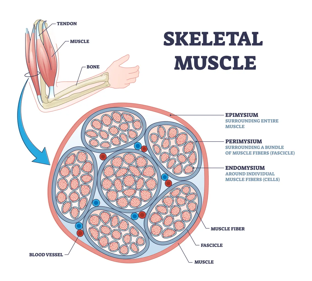
Muscle fibre plasticity
Our muscles are plastic.
By this, of course, I do not mean that our muscles are made up of PVC (although, like synthetic plastics, the contractile proteins that make up muscle fibres, myosin and actin, are also polymers, composed of chains of smaller repeating units). Rather, being “plastic” means that the composition of a particular skeletal muscle, namely its relative proportions of fast- and slow-twitch muscle fibres, changes in response to how that muscle is used.
From an evolutionary perspective, it’s easy to see why this plasticity of muscle fibre composition is useful.
Say your ancestor found herself in an environment that required her to run for prolonged periods to hunt deer. Running, which involves repeatedly exerting force on the ground to move forwards, employs slow-twitch (Type I) fibres, which are able to contract repeatedly for long periods without fatiguing.
Over time, by increasing the proportion of slow-twitch (Type I) fibres in her leg muscles, your ancestor will become better adapted to running, more effective at hunting prey, and, overall, more suited to the demands of her particular environment. Accordingly, this would have increased her chances of survival and successfully reproducing.
Now, suppose that your ancestor migrates to a new terrain that, rather than prolonged running, necessitates her having to regularly move heavy rocks in order to access water and berries. This new activity involves heavy mechanical loading of upper body muscles and makes greater use of fast-twitch (Type II) muscle fibres. These are capable of generating much higher forces than slow-twitch fibres (but fatigue more easily). By increasing the proportion of fast-twitch fibres in her upper body muscles, your ancestor is able to adapt to her new environment, again increasing her survival and reproductive chances.
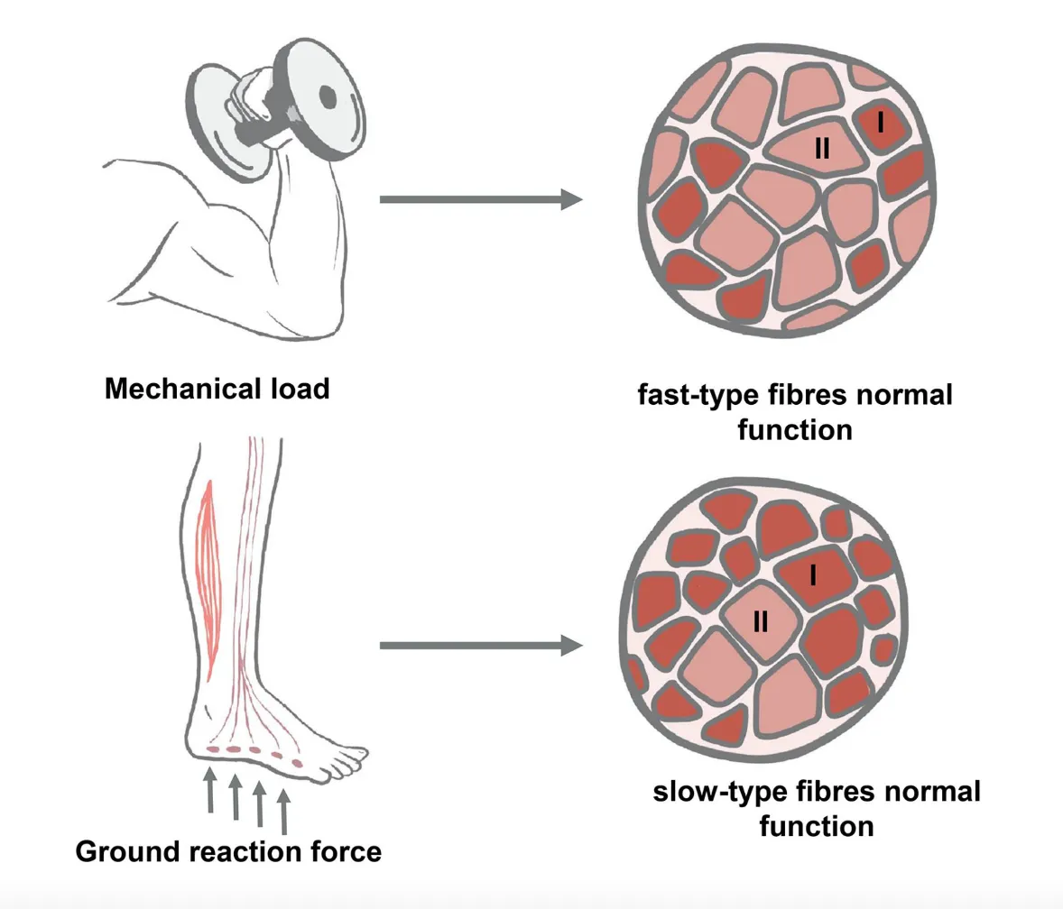
Heavy mechanical loading, as in resistance exercise, increases the proportion of fast-twitch (Type II) muscle fibres. By contrast, endurance activities such as running increase a muscle’s proportion of slow-twitch (Type I) fibres.
Consider, though, that the number of skeletal muscle fibres we have is thought to be largely fixed from birth. Studies in other mammals suggest that we produce new muscle fibres while developing in utero; but this process doesn’t take place significantly after we’re born. Clearly, our muscles become bigger and stronger as we grow from babies into adults, but this is predominantly due to enlargement of the cross-sectional area of individual muscle fibres (hypertrophy) rather than increases in the number of muscle fibres.
Given this constraint on our total number of muscle fibres and the fact that we cannot simply add more slow- and fast-twitch fibres to a muscle per se, how do we change our muscle fibre composition?
The answer is that muscle fibres can transition: they can change from one type into another.
In this respect, burgeoning evidence suggests that fast-twitch (Type II) fibres can convert into slow-twitch (Type I) fibres, and vice versa. By interconverting different fibre types in a given muscle, we end up altering that muscle’s proportions of slow- and fast-twitch fibres. Converting slow-twitch into fast-twitch fibres in our biceps, for instance, will increase its relative proportion of fast-twitch fibres (while simultaneously decreasing the proportion of slow-twitch fibres). It is such transitions between muscle fibre types that underlies the plasticity of our muscles.
As we’ll describe in greater detail below, studies demonstrate that specific modes of exercise can stimulate particular muscle type transitions. Before we learn about how muscle fibres transition from one type into another, however, we need to establish what these different muscle types are.
Different muscle fibre types
So far we’ve implicitly distinguished between two main types of muscle fibre based on how quickly they contract: slow-twitch (Type I) and fast-twitch (Type II).
Rather than muscle fibres falling into two distinct camps, a more accurate picture is that our muscle fibres lie on a continuum from slow to fast contraction speeds. Think ‘shades of grey’ rather than ‘black and white’. This, of course, raises the question: “How do we decide which fibres are ‘slow-twitch’ and which are ‘fast-twitch’?”
Naturally, carving up any continuum into discrete categories is fraught with difficulty. (Consider, for example, the pressing metaphysical question of “At what point does bread become toast?”). Alas, if we probe deeper, it appears that one of the reasons muscle fibres contract at different speeds is due to differences in the form of contractile proteins they produce. We can use these differences in protein form to help us classify muscle fibres into discrete groups.
MHC types
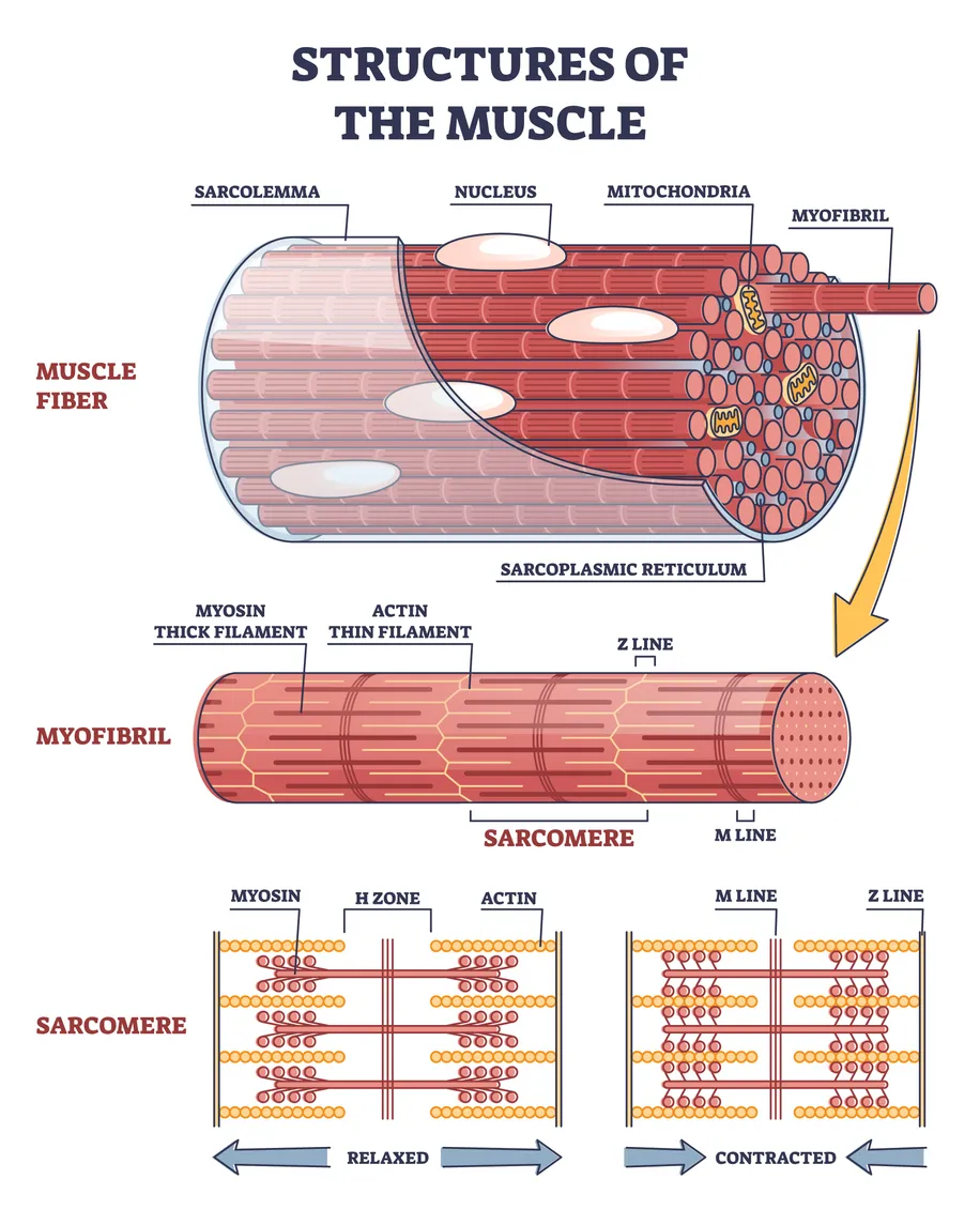
As mentioned earlier, one of the major contractile proteins in our muscle is myosin. In very simple terms, myosin acts like a motor in sarcomeres: the basic contracting units that make up a muscle fibre.
Thick filaments made out of myosin use chemical energy (in the form of ATP) to quickly change shape and pull other thin filaments (made out of actin) together in what is known as a “power stroke.” It is this action of pulling filaments so that they slide over one another that ultimately leads to contraction of muscle fibres (see image below).
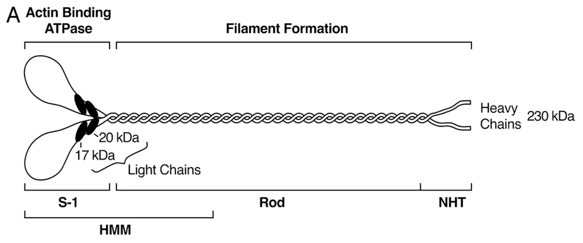
An individual myosin molecule is composed of two heavy chains (MHC) and four light chains (MLC) (see diagram below). These chains come in subtly different forms, known as isoforms. In humans, the main three isoforms of the myosin heavy chain (MHC) are:
- MHC Type I
- MHC Type IIa
- MHC Type IIx.
It turns out that the MHC isoform a muscle fibre predominantly produces (or ‘expresses’) strongly influences how quickly that muscle fibre contracts, how much force it can generate, and how it derives chemical energy in order to contract.
Muscle fibres producing MHC Type I tend to contract more slowly and cannot generate high forces. Reliant on aerobic respiration (i.e using oxygen) to generate energy, however, these fibres can contract repeatedly for long periods and are highly resistant to fatigue. Accordingly, these fibres are useful for endurance activities. We call these MHC Type I-producing fibres slow-twitch, slow-oxidative fibres, or simply, Type I fibres.
MHC Type IIa-producing fibres are called fast oxidative glycolytic fibres, or simply Type IIa fibres. They can contract more quickly and generate higher forces than Type I fibres, but fatigue more quickly. By virtue of their greater force production, Type IIa fibres are useful for strength and power activities. They can use either aerobic or anaerobic (i.e without oxygen) respiration to generate energy.
The muscle fibres that contract the fastest and generate the highest forces are those producing MHC Type IIx. These fast glycolytic fibres or simply, Type IIx fibres, are fully-reliant on anaerobic respiration for energy and, as such, fatigue very quickly. Type IIx fibres are particularly useful for power activities involving explosive movements.
The graph below nicely illustrates the differences in contraction velocity and maximum force between the different fibre types based on the MHC isoform they produce.

Some muscle fibres, known as hybrid muscle fibres, also produce more than one MHC isoform. For example, Type I/IIa fibres produce a mixture of both MHC Type I and Type IIa.
Hybrid fibres can be thought of as intermediate fibres, combining the properties of different MHC types. Accordingly, a Type I/IIa hybrid fibre is expected to contract more quickly and forcefully than a pure Type I muscle fibre, but less quickly than a pure Type IIa fibre. Hybrid fibres may also be conceived of as an ‘in-between stage’ as muscle fibres transition from type into another - more on that later.
In summary, carving up the continuum of slow to fast muscle fibres by what MHC type each fibre produces gives us the following types:
- Type I (slow-oxidative) fibres
- Type IIa (fast oxidative glycolytic) fibres
- Type IIx (fast glycolytic) fibres
- Hybrid fibres (e.g. Type I/IIa, that combine different MHC types).
(To quickly revert back to the slow- vs fast-twitch distinction: Type I fibres are slow-twitch and Type IIa and IIx fibres can be grouped together as fast-twitch.)
Now we’ve established the different muscle fibre types, let’s find out what happens when one type changes or transitions into another.
Muscle fibre transitions
When we exercise, the patterns of electrical activity in nerves that supply our muscles change. Similarly, our muscles are subject to various mechanical and metabolic stressors (such as the physical load of a heavy weight or the build-up of lactate and H+ ions that accompany anaerobic respiration).
These exercise-induced changes in electrical and molecular activity stimulate various signalling pathways that cause muscle fibres to start producing different MHC isoforms.
For example, in response to the prolonged, repeated muscle contractions that characterise endurance running, a Type IIa muscle fibre, which ordinarily produces MHC Type IIa, might start producing MHC Type I. In doing so, it transitions from a Type IIa fibre into a hybrid Type IIa/I fibre. With further training, this hybrid Type IIa/I fibre may then start only producing MHC Type I, thus becoming a pure Type I fibre.
What was once a fast-twitch, easily fatigable Type IIa fibre has now, after weeks of endurance training, transformed into a slow-twitch, fatigue resistant Type I fibre. As similar transitions occur in other muscle fibres for a given muscle, the proportion of Type I fibres in that muscle will increase. At the same time, the proportion of Type IIa fibres will decrease.
Scientists are able to visualise these changes in muscle fibre type proportions by taking biopsies of muscle tissue, staining them using antibodies targeted to the different MHC isoforms, and then studying them under a microscope.
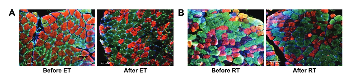
The images above, for example, are images taken from biopsies of the thigh muscles (vastus lateralis) of two different COPD patients before and after exercise training. The left-hand image (A) shows the impact of endurance training (ET), while the right-hand image (B) shows that of resistance training (RT). As we can clearly see, the proportion of fibres producing MHC Type I, which are stained green, has significantly increased following endurance training. Conversely, resistance training has increased the proportion of muscle fibres producing MHC Type IIa, which are stained red.
Due to the varying demands they place on muscles and, correspondingly, the different signalling pathways they activate, endurance, resistance, and sprint training elicit markedly different muscle fibre type transitions and changes in muscle fibre proportions.
Let’s take a look at what each exercise mode does.
Resistance training and muscle fibre type transitions: IIx → IIa
A large body of research suggests that resistance (strength) training tends to cause fast glycolytic Type IIx and hybrid Type IIa/IIx fibres to transition into pure fast oxidative glycolytic Type IIa fibres.
Pumping iron and hitting the weight rack will therefore increase your proportions of pure Type IIa fibres in whichever muscles you exercise, while decreasing the proportion of Type IIx fibres. Illustrating this, the graph below comes from a 2003 study by Y. Liu and colleagues, in which half of subjects completed 6 weeks of resistance training, performing bench presses at 3-RM (3 rep max) for three days a week.
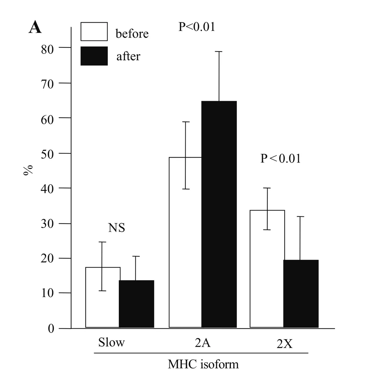
As can be seen by comparing the different black and white bars, the proportion of Type IIa fibres in the triceps brachii muscle shot up significantly from 49.4% to 66.7% after training, while the proportion of Type IIx fibres dropped from 33.4% to 19.5%.
But, what happened to slow-twitch Type I fibres? As shown in the above graph, the proportion of these stayed the same. This is in line with several other studies, which suggest that resistance training doesn’t significantly elicit Type I into Type IIa muscle fibre transitions.
That is not to say it is impossible to convert Type I into Type IIa fibres.
Returning to the aforementioned Liu et al study; while one half of subjects purely performed bench presses over the 6-week training period, the other half incorporated ballistic and plyometric exercises into their training regime. For two of the exercise days, they performed bench press throws and plyo push-ups - both explosive movements that employ the high-velocity, high-force contractions of Type IIx muscle fibres.
So, what happened to the muscle fibre composition of this group?
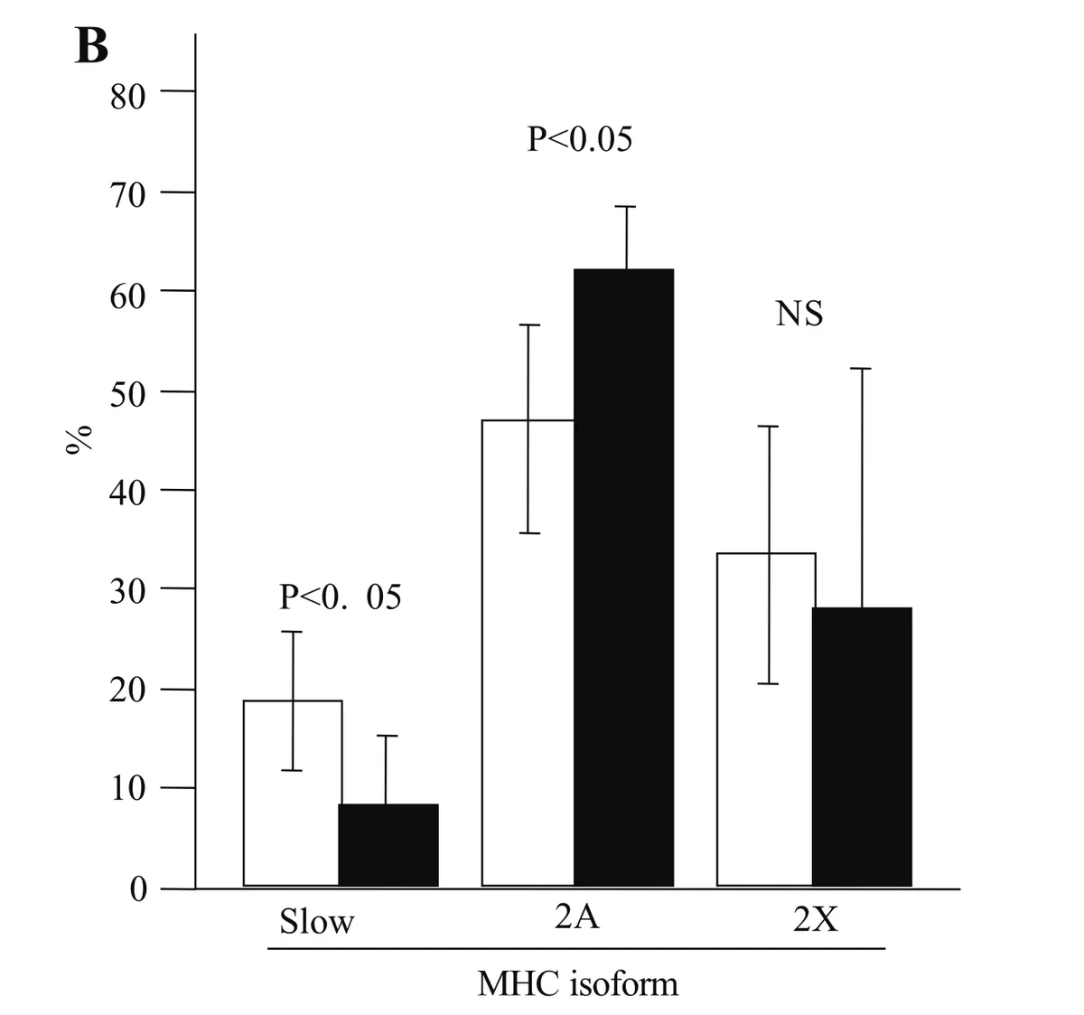
As with those performing bench presses only, subjects performing a combination of bench presses and explosive exercises increased their proportion of Type IIa fibres. This higher proportion, however, didn’t come at the expense of Type IIx fibres, which stayed at roughly the same proportion (NS - indicates a non-significant change in the above graph). Rather, the proportion of slow-twitch Type I fibres decreased significantly from 18.2% to 9.2%, suggesting it was these (rather than Type IIx fibres) that transitioned into Type IIa fibres.
These findings tell us that incorporating explosive, high-velocity movements into resistance training can preserve Type IIx fibres, while promoting the transition of Type I into Type IIa fibres.
Sprint training and muscle fibre type transitions: I → IIa
As with resistance training, sprint training is also demonstrated to increase the proportion of fast oxidative glycolytic Type IIa fibres.
Unlike conventional resistance training, however, which typically involves higher loads and lower muscle contraction speeds, sprint training relies on faster muscle contractions and may more strongly elicit Type I into Type IIa muscle fibre transitions.
The image below shows the biopsies (taken from the vastus lateralis thigh muscle) of a subject before (PRE), two weeks into (MID), and after (POST) a 6-week sprint-interval training programme.
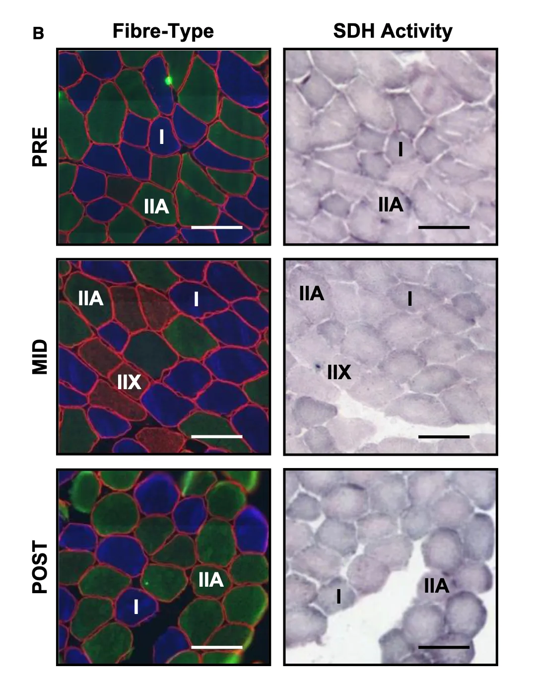
As can be seen in the left-hand column of images, the proportion of Type IIa muscle fibres, stained green, increased after sprint training, while the proportion of Type I fibres, stained blue, decreased. This suggests sprint training stimulated Type I fibres to transition into Type IIa fibres.
Notice also that at two weeks into training, the number of Type IIx fibres, stained red, increased. By the end of training, however, this proportion had dropped back. Changes in proportions of Type IIx fibres in response to training are generally less well understood, partly because pure Type IIx fibres are the least common of muscle fibre types, thought to make up less than 2% of all muscle fibres.
Intuitively, we might expect sprint training to increase our proportion of Type IIx fibres, as these are useful for the powerful, high-velocity contractions that support sprinting. Contrary to this hypothesis, a few studies have found that sprint training actually causes Type IIx and hybrid IIa/IIx fibres to transition into Type IIa fibres, thereby decreasing the proportion of Type IIx fibres.
By contrast, thigh muscle biopsies of elite sprinters have found that they have considerably higher proportions of pure Type IIx fibres (around 6% according to some estimates). For one former world record holder of the 110m hurdles, this figure was even as high as 24%! It is therefore possible that, through years of intense training, sprinters are able to convert other fibre types (such as hybrid IIa/IIx fibres) into pure Type IIx fibres, thereby boosting their proportion of pure Type IIx fibres. Of course, it is also possible that elite sprinters have simply been genetically-endowed with a greater baseline proportion of Type IIx fibres.
Endurance training and muscle fibre type transitions: Type IIa → Type I
Unsurprisingly, endurance training elevates the proportions of slow-twitch Type I oxidative fibres, which are adapted for repeated contractions over longer time periods.
This shift in proportions likely results from the transition of Type IIa and hybrid Type I/IIa fibres into pure Type I fibres.
On this note, a study of amateur runners undergoing 16 weeks of marathon training found that the proportion of Type I fibres in their vastus lateralis thigh muscle increased from 42.6% to 48.6%, while the proportion of Type IIa fibres dropped from 40.1% to 35.8%.
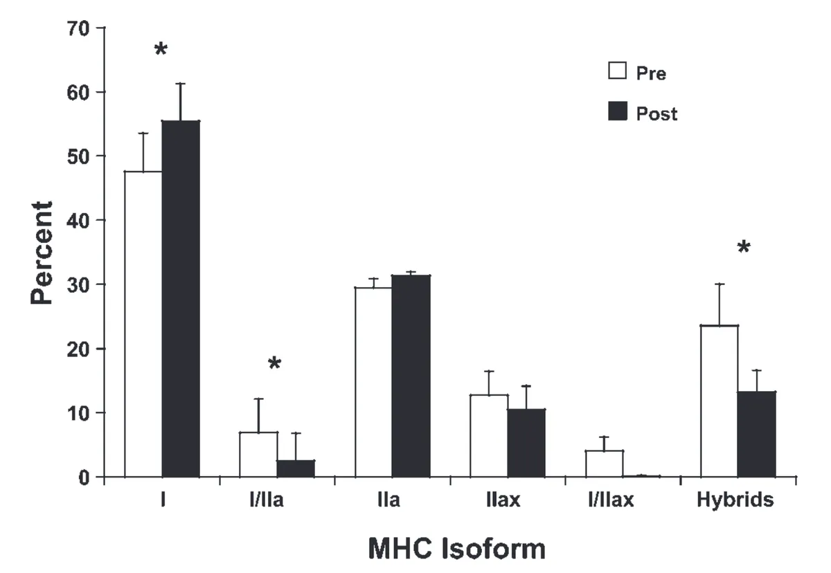
Another study of marathon training found that as well as elevating the proportion of Type I fibres in the gastrocnemius muscle (one of two major calf muscles), the percentage of hybrid Type I/IIa fibres fell (see graph above). This intimates that these hybrid fibres potentially transitioned into pure Type I fibres.
Putting things in context
We’ve discussed how you can stimulate muscle fibre type transitions and change your muscle fibre composition with various forms of exercise. Does this mean you can completely repopulate your thighs with fast-twitch Type IIa fibres?
No. Firstly, not all of our muscles are the same. The baseline muscle fibre composition of various skeletal muscles differs according to anatomical site. For instance, the extensor digitorum longus muscle, which runs along the front of our lower leg, is predominantly composed of fast-twitch Type IIa and Type IIx fibres. By contrast, the soleus, one of our two calf muscles, has a much larger proportion of slow-twitch Type I fibres.
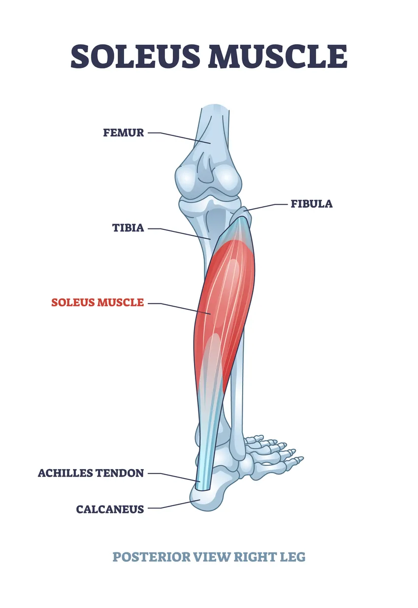
Secondly, fibres in individual muscles have varying propensities to transition in response to exercise. Type I muscle fibres in the soleus, for example, may be less likely to convert into Type IIa and other fibre types. Interestingly, this may reflect how individual muscles have been shaped by evolution for different movements. The soleus inserts into your Achilles tendon and helps plantar-flex your ankle (i.e. move it downwards towards the ground), a movement important for walking and running. It seems sensible that a muscle designed for walking and running remains laden with Type I muscle fibres that can contract repeatedly without fatiguing.
Consonant with this, the proportions of muscle fibres we’re born with, and our potential to alter a particular muscle’s composition with different forms of exercise, are likely strongly influenced by our genetics. On this note, a study of twins found that 45% of the difference in muscle fibre composition was due to genetic factors. While some of us are genetically inclined to be fast-twitch dominant sprinters, others of us, with a genetic preponderance of slow-twitch Type I fibres, are more natural marathoners.
Our muscles may be plastic, but this plasticity is constrained by our genes.
Image references
Sharlo, K., Tyganov, S. A., Tomilovskaya, E., Popov, D. V., Saveko, A. A., & Shenkman, B. S. (2022). Effects of various muscle disuse states and countermeasures on muscle molecular signaling. International Journal of Molecular Sciences, 23(1), 468.
Ma, X., & Adelstein, R. S. (2014). The role of vertebrate nonmuscle Myosin II in development and human disease. Bioarchitecture, 4(3), 88-102.
Polla, B., D’antona, G., Bottinelli, R., & Reggiani, C. (2004). Respiratory muscle fibres: specialisation and plasticity. Thorax, 59(9), 808-817.
Iepsen, U. W., Munch, G. D. W., Rugbjerg, M., Rinnov, A. R., Zacho, M., Mortensen, S. P., ... & Thaning, P. (2016). Effect of endurance versus resistance training on quadriceps muscle dysfunction in COPD: a pilot study. International journal of chronic obstructive pulmonary disease, 2659-2669.
Liu, Y., Schlumberger, A., Wirth, K., Schmidtbleicher, D., & Steinacker, J. M. (2003). Different effects on human skeletal myosin heavy chain isoform expression: strength vs. combination training. Journal of Applied Physiology, 94(6), 2282-2288.
Edgett, B. A., Bonafiglia, J. T., Baechler, B. L., Quadrilatero, J., & Gurd, B. J. (2016). The effect of acute and chronic sprint‐interval training on LRP 130, SIRT 3, and PGC‐1α expression in human skeletal muscle. Physiological reports, 4(17), e12879.
Trappe, S., Harber, M., Creer, A., Gallagher, P., Slivka, D., Minchev, K., & Whitsett, D. (2006). Single muscle fiber adaptations with marathon training. Journal of applied physiology, 101(3), 721-727.